Aragona Group - Tissue Architecture
The Aragona group aims to understand how neighbouring cells coordinate their decisions to build tissues with specialized structure and function. In particular, they want to investigate how tissue architecture is maintained and how mechanical cues affect gene transcription, stem cells dynamics and fate decisions.
The questions the group wants to address include how tissues remodel their cellular division and cell-fate patterns, and how the cell-cell and cell-matrix connections are adjusted to coordinate biological function and collectively react to external stress with the activation of specific transcription factor programs. They are investigating these biological questions in tissues that are naturally subjected to different mechanical perturbations such as the skin and the urinary tract. Understanding these fundamental principles will improve our basic understanding and have a high impact in the development of new regenerative medicine tools.
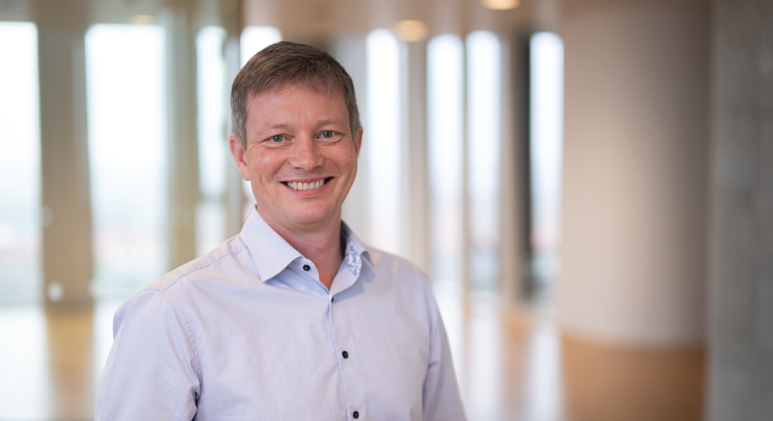
Associate Professor Mariaceleste Aragona
mariaceleste.aragona@sund.ku.dk
CURIS
Google Scholar profile
ORCID profile
The functionality of each organ in our body is determined by the structure and correct organisation of each tissue component. Tissue architecture is ensured by a perfect equilibrium between each individual cell, its neighbours and the physical environment. In particular, this is tightly regulated in epithelia that are constantly renewed through our lives thanks to stem cells and progenitors able to regenerate entire tissues. Stem cells have the capacity to renew and differentiate in other cells types and the ability to sense and cope with different external cues. Specifically, we focus our interest into the mechanobiology field that studies the set of mechanisms converting physical cues into chemical signals by a process called mechanotransduction.
The questions we want to address include how tissues remodel their cellular division and cell-fate patterns, and how the cell-cell and cell-matrix connections are adjusted to coordinate biological function and collectively react to external stress with the activation of specific transcription factor programs. We are investigating these biological questions in tissues that are naturally subjected to different mechanical perturbations such as the skin and the urinary tract. Understanding these fundamental principles will improve our basic understanding and have a high impact in the development of new regenerative medicine tools.
Using lineage tracing in mouse models, embryo tissue explants for live imaging during organ formation, transcriptomic and chromatin profiling at the single cell level, we study how mechanical stimuli are integrated by the cells and translated in signalling pathways that, providing positional information for cell fate decisions, directly shape tissue architecture.
Mechanisms regulating the response to stretching in the skin
The capacity of the skin to expand as a reaction to stretching is a property commonly exploited in plastic surgery to generate an excess of skin that can be used to repair birth defects, damaged tissues, scars and for breast reconstruction after mastectomy. Although this technique has been used for decades in reconstructive surgery, little is known about the cellular and molecular responses of cells to tissue stretching. For these reasons, the skin is a perfect tissue to study and model the effects of mechanical stimuli on cell behaviour in a clinically relevant context. We developed a mouse model in which the temporal consequences of stretching the skin epidermis can be studied in vivo. We discovered that stretching induces skin expansion by creating a transient bias in the renewal activity of epidermal stem cells, while a second subpopulation of basal progenitors remains committed to differentiation. By transcriptional and chromatin profiling and functional assays, we identified how cell states and gene regulatory networks are modulated by stretching in the epidermis. We are currently studying how the other skin compartments, mainly in the dermis, maintain themselves under mechanical stress, as well as how they coordinate their own homeostatic behavior with the different neighboring cell types.
A mouse model to study mechanotransduction in the skin. Several cell types constitute and maintain the barrier function of the skin. We are studying how they all ensure proper tissue architecture in response to mechanical cues such as mechanical stretching
Principles of cellular urinary tract homeostasis
It has become increasingly clear that not only stretching and compression are important physical cues that shape epithelia, but that also more broadly acting forces like fluid flow and hydrostatic pressure play substantial roles during morphogenesis and homeostasis. As complementary paradigm, we are studying the epithelium of the urinary tract that is under frequent mechanical loading and unloading due to the urine flow, providing a model in which we can interrogate the effect of fluid mechanics on epithelia biology. Additionally, from these body compartments, with the urine flow, we lose cells that need to be constantly replaced. However, it is still unknown whether this tissue is maintained by a single equipotent population of cells or by a defined cluster of stem cells and which degree of plasticity characterises these cells. By single-cell sequencing, lineage tracing and clonal analysis, we are studying the hierarchical organisation and proliferation dynamics of cell lineages within the ureters and urethra epithelia.
Urinary tract tubulogenesis and development
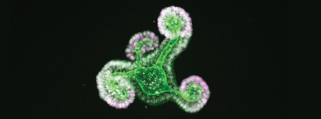
With the tools and knowledge acquired by studying the urethral epithelium during homeostasis, we are interrogating the cellular mechanism of lumen formation during embryonic development. We are studying which are the driving forces that, from a single homogeneous group of cells, specify and pattern the different cell types to generate the adult tissue architecture. By culturing ex vivo mouse embryonic explants we are investigating the cellular dynamics that lead to tubulogenesis from a spatial and temporal perspective. To understand whether and how fluid mechanics affect the generation of the organ, we are correlating changes in cell-fates and in the remodelling of the actomyosin cytoskeleton with the measurements of the hydrostatic pressure and urine flow. By studying these processes, we will be able to shed light on the molecular mechanisms that could be responsible for congenital anomalies and disorders affecting the urinary tract.
Mechanotransduction in the urethral epithelium
To gain further insights in the molecular characterisation of this biological system, we will implement currently available 3D organotypic cultures of human urinary tract tissue, and use them to analyse the cells dynamics in response to the pressure and shear stress generated by the urine flow. By live imaging on organoid cultures we will track the different cell fates, we will analyse the molecular mechanisms and the gene regulatory networks underlying cell behaviors, and we will describe in which specific cells different signaling pathways are activated or repressed.
Globally, we aim to understand how mechanical cues, morphogens, cell-cell interaction and competition, as well as other important determinants of the cell microenvironment and niche, locally regulate transcription to shape tissue architecture and how this impacts stem cell physiology and pathology. Our ultimate goal is to provide deep knowledge on how tissues are naturally built, in order to direct new approaches for regenerative and reconstructive therapies.
Nguyen, T. M., and Aragona, M. (2021). Stress-responsive transcription factors train stem cells to remember. Cell Stem Cell, 28(10), 1679–1680. doi:10.1016/j.stem.2021.09.005.
Nguyen, T. M., and Aragona, M. (2021). Regulation of tissue architecture and stem cell dynamics to sustain homeostasis and repair in the skin epidermis. Seminars in Cell and Developmental Biology, S1084-9521(21)00243-3. doi: 10.1016/j.semcdb.2021.09.008.
Aragona, M., Sifrim, A., Malfait, M., Song, Y., Van Herck, J., Dekoninck, S., Gargouri, S., Lapouge, G., Swedlund, B., Dubois, C., Baatsen, P., Vints, K., Han, S., Tissir, F., Voet, T., Simons, B., and Blanpain, C. (2020) Mechanisms of stretch-mediated skin expansion at single-cell resolution. Nature, 584(7820), 268-273. doi: 10.1038/s41586-020-2555-7.
Dekoninck, S.#, Hannezo. E.#, Sifrim, A., Miroshnikova, Y. A., Aragona, M., Malfait, M., Gargouri, S., de Neunheuser, C., Dubois, C., Voet, T., Wickström, S. A., Simons, B. D., and Blanpain, C.°(2020). Defining the Design Principles of Skin Epidermis Postnatal Growth. Cell, 604-620. doi: 10.1016/j.cell.2020.03.015.
Aragona, M.#, Dekoninck, S.#, Rulands, S., Lenglez, S., Mascré, G., Simons, B. D., and Blanpain, C. (2017). Defining stem cell dynamics and migration during wound healing in mouse skin epidermis. Nature Communications, 8(1), 1-14. doi: 10.1038/ncomms14684.
Aragona, M. and Blanpain, C. (2017). Gene therapy: Transgenic stem cells replace skin. Nature, 551(7680), 306-307. doi: 10.1038/nature24753.
Enzo, E., Santinon, G., Pocaterra, A., Aragona, M., Bresolin, S., Forcato, M., Grifoni, D., Pession, A., Zanconato, F., Guzzo, G., Bicciato, S., and Dupont, S. (2015). Aerobic glycolysis tunes YAP/TAZ transcriptional activity. The EMBO Journal, 34, 1349-1370. doi: 10.15252/embj.201490379.
Aragona, M., Panciera, T., Manfrin, A., Giulitti, S., Michielin, F., Elvassore, N., Dupont, S., and Piccolo, S. (2013). A mechanical checkpoint controls multicellular growth through YAP/TAZ regulation by actin-processing factors. Cell, 154, 1047-1059. doi: 10.1016/j.cell.2013.07.042.
Dupont, S., Morsut, L, Aragona, M., Enzo, E., Giulitti, S., Cordenonsi, M., Zanconato, F., Le Digabel, J., Forcato, M., Bicciato, S., Elvassore, N., and Piccolo, S. (2011). Role of YAP/TAZ in mechanotransduction. Nature, 474(7350), 179-183. doi: 10.1038/nature10137.
# equal contributions
Research Profile
Mariaceleste background covers many aspects of modern developmental and molecular biology. She obtained her Master's degree in Medical Biotechnology from the University of Padova in Italy. During her PhD, under the supervision of Professor Stefano Piccolo, she learned to develop, optimise and standardise new bioassays, from early xenopus and mouse embryology to stem cell differentiation, acquiring as well the competence to uncover the crosstalk within different molecular signalling pathways. In this period, she became fascinated by the mechanobiology field while researching transcription factors able to read the mechanical stimuli instructed on cells by the extracellular matrix (ECM) composition and shape, and translate them into gene transcription and cell functions. With her PhD thesis, in collaboration with Professor Sirio Dupont, she greatly contributed to the identification of YAP and TAZ (the downstream factors of the Hippo pathway) as sensors and mediators of mechanical cues. During a short post-doc, carried out still in Padova, she identified F-actin capping and severing proteins Cofilin, CapZ and Gelsolin as inhibitors of YAP and TAZ activity. In this study, she showed that the architectural features of an epithelial sheet – 3D shape and rigidity of its surrounding ECM – define the locations where cell proliferation is allowed, by generating patterns of mechanical forces that spatially regulate the activity of YAP and TAZ.
In 2014, Mariaceleste joined the team of Professor Cédric Blanpain at Université Libre de Bruxelles in Belgium, where she increased her knowledge on epithelia biology and learned how to study stem cell fate decision using a combination of lineage tracing, proliferation kinetics, molecular profiling and mathematical modelling, in close collaboration with Professor Benjamin Simons at Cambridge University, UK. She uncovered how different stem cells populations in the skin epidermis balance proliferation, migration and differentiation during wound healing. Thanks to a prestigious fellowship of the Human Frontier Science Program (HFSP) and to the Belgian Fund for the Scientific Research (FNRS), she studied how mechanical stretching impacts skin stem cell behaviour. By combining clonal analysis, single cell RNA sequencing and chromatin profiling, she dissected step-by-step the clonal dynamics and molecular mechanisms by which epidermal stem cells respond to mechanical forces in vivo.
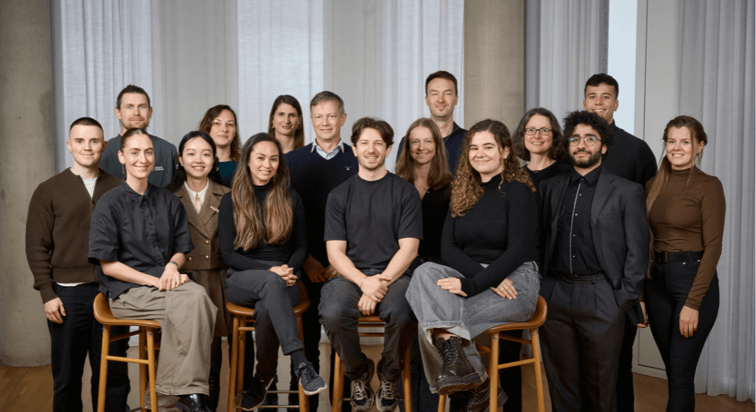
The Aragona Laboratory at reNEW is now focused on understanding how mechanical cues shape tissue architecture and change epithelial stem cells dynamics during homeostasis and organ growth. The goal of the lab is also to establish a collaborative environment in which creativity is supported, and in which each person contributes to the success of each other individual and common ambitions.
Curriculum Vitae
January 2022: Associate Professor, Group Leader
University of Copenhagen, Denmark
Novo Nordisk Foundation Center for Stem Cell Medicine, reNEW
September 2020: Associate Professor, Group Leader
University of Copenhagen, Denmark
Novo Nordisk Foundation Center for Stem Cell Biology, DanStem
May 2014 – July 2020: Post-Doctoral Training
Université Libre de Bruxelles, Belgium
Advisor: Professor Cédric Blanpain
April 2012 – April 2014: Post-Doctoral Training
University of Padova, Italy
Advisor: Professor Stefano Piccolo
January 2009 – April 2012: Doctorate in Biomedicine
University of Padova, Italy
Advisor: Professor Stefano Piccolo
September 2006 - October 2008: Master in Medical Biotechnology
University of Padova, Italy
Advisor: Dott. Paola Brun
September 2003 – September 2006: Bachelor in Biotechnology
University of Padova, Italy
Advisor: Professor Daniela Danieli
Mariaceleste Aragona: The architecture of tissues and organs
Staff List - Aragona Group
Name | Title | Job responsibilities | Phone | |
---|---|---|---|---|
Search in Name | Search in Title | Search in Job responsibilities | Search in Phone | |
Aguilera, Caroline | PhD Fellow | PhD Fellow | ||
Apresentação de Almeida, João Tiago | External | |||
Aragona, Mariaceleste | Associate Professor | |||
Axelsen, Oscar | External | |||
Bargsted Elgueda, Leslie Ann | Postdoc | +4535328706 | ||
Kaklamanou, Ioanna | PhD Fellow | PhD Fellow | ||
Magnussen, Michael James | Postdoc | +4535326281 | ||
Miskovic Krivokapic, Jelena | Academic Research Officer | |||
Teixeira, Beatriz | Research Assistant | +4535328364 |